


Knee arthropathy, the most common site of degenerative dystrophic disease, is characterized by the progressive destruction of cartilage followed by changes in the articular surface, with pain and decreased mobility.
The disease is more likely to affect women over the age of 40, especially those who are overweight and have varicose veins in the lower extremities.
The knee joint consists of three compartments:
- medial tibiofemoral;
- lateral tibiofemoral;
- Suprapatellar - femur.
These compartments can be affected by deforming osteoarthritis (DOA) individually or in any combination. 75% of knee arthropathy cases are destruction of the medial tibiofemoral compartment (under 2-3 times body weight during exercise).
In younger patients, only one joint is more commonly destroyed - the right or left side (right or left arthropathy).
Causes of knee DOA
Several factors may simultaneously be involved in the development of degenerative cartilage changes:
- Mechanical overload of the knee joint (in some specialties, sports) with minimal cartilage trauma;
- Injury, consequences of surgical intervention (meniscectomy);
- Inflammatory disease of the knee joint (arthritis);
- anatomical inconsistencies of the articular surfaces (dysplasia);
- Violation of statics (flat feet, curvature of the spine);
- Chronic hemoarthritis (hemosynovia);
- metabolic pathology (gout, hemochromatosis, chondrocalcinosis);
- being overweight;
- Violation of the blood supply to the bones;
- osteodystrophy (Paget's disease);
- Neurological disorders, loss of sensation in the extremities;
- endocrine disorders (acromegaly, diabetes, amenorrhea, hyperparathyroidism);
- Genetic susceptibility (a general form of osteoarthritis);
- Violates the synthesis of type II collagen.
But in 40% of cases, the underlying cause of the disease (primary arthropathy) cannot be identified.
Pathogenesis of knee joint disease
Early stage
In the initial stages of the disease, cartilage metabolic processes are disturbed. Synthesis and mass reduction of proteoglycans, the main building blocks of cartilage tissue responsible for the structural stability of the collagen network.
As a result, chondroitin sulfate, keratin, and hyaluronic acid are washed out of the mesh, and structurally defective proteoglycans can no longer retain water. It is absorbed into collagen, and its swollen fibers cause the cartilage to become less resistant to pressure.
In the synovial cavity, pro-inflammatory substances accumulate, under their influence the cartilage is destroyed more rapidly. Development of capsular fibrosis. Changes in the composition of synovial fluid make it difficult to deliver nutrients to the cartilage and impair the sliding of articular surfaces during movement.
pathological progression
In the future, the cartilage will gradually thin and become rough, with cracks appearing throughout the thickness. The epiphysis of the bone is subjected to an increased load, which triggers osteosclerosis and compensatory proliferation of bone tissue (osteophytes).
This response of the body is designed to increase the area of the articular surfaces and redistribute the load. But the presence of osteophytes can increase discomfort, deformity, and further limit mobility of the limb.
Microfractures form in the thickness of the bone, which can damage blood vessels and cause high pressure within the bone. In the final stages of osteoarthritis, the articular surfaces are completely exposed, deformed, and limb movement is severely restricted.
Symptoms of Knee Arthropathy
Knee arthropathy is characterized by a chronic, slowly progressive course (months, years). The clinic developed gradually, with no apparent deterioration. Patients cannot remember exactly when the first symptoms appeared.
Clinical manifestations of knee joint disease:
- pain. Diffuse, transient at first (standing for long periods of time, going up stairs), as osteoarthritis progresses, the pain becomes localized (front and inner surface of the knee) and its intensity increases;
- Local palpation sensitivity. Mostly on the medial side of the knee joint along the edge of the joint space;
- crunch. In the first stage it may be inaudible, in the second stage it accompanies all movements;
- The volume increases and the knee deforms. A person's limbs are O-shaped (clearly visible even in the photo) due to weakening of the lateral ligaments;
- Liquidity restrictions. At first, there is difficulty in bending the knee, and later - stretching.
Causes of DOA pain:
- Mechanical friction of damaged articular surfaces;
- Increased intraosseous pressure, venous congestion;
- the addition of synovitis;
- Changes in the tissues around the joint (stretching of capsules, ligaments, tendons);
- Periosteal thickening;
- Dystrophy of adjacent muscles;
- fibromyalgia;
- Compression of nerve endings.
In contrast to hip arthropathy, knee DOA may exhibit spontaneous resolution of symptoms.
The clinical presentation of knee arthropathy depends on the stage:
| feature | i stage | Phase II | The third phase |
|---|---|---|---|
| pain | Short, occurs more often when the knee is extended (standing for long periods of time, walking up stairs) | Moderate, disappears after a night of rest | pronounced pronounced, disturbing even at night |
| Movement restrictions | Invisible | Restricted extension, mild limp | Persistent flexor and extensor contractures, lameness |
| crunch | no | Palpation sensation during exercise | remote crunch |
| deformation | Missing | Slight forward movement of limb axis, muscle atrophy | Valgus or varus deformity. Joint instability, thigh muscle atrophy |
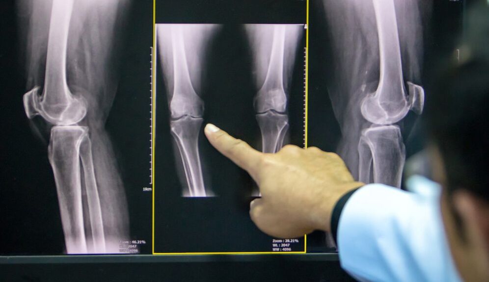
| X-ray pictures | Slight narrowing of the joint space, initial signs of subchondral bone sclerosis | The joint space is reduced by more than 50%, and osteophytes appear | Almost complete absence of joint space, marked deformation and hardening of the articular surface, subchondral necrotic areas of bone, osteoporosis |
A common complication of knee arthropathy is secondary reactive synovitis, characterized by the following symptoms:
- growing pains;
- puffiness;
- Fluid enters the synovial cavity;
- Elevated skin temperature.
Less common and more dangerous complications include: joint obstruction, osteonecrosis of the femoral condyle, patellar subluxation, and spontaneous hemoarthrosis.
Diagnosis of knee DOA
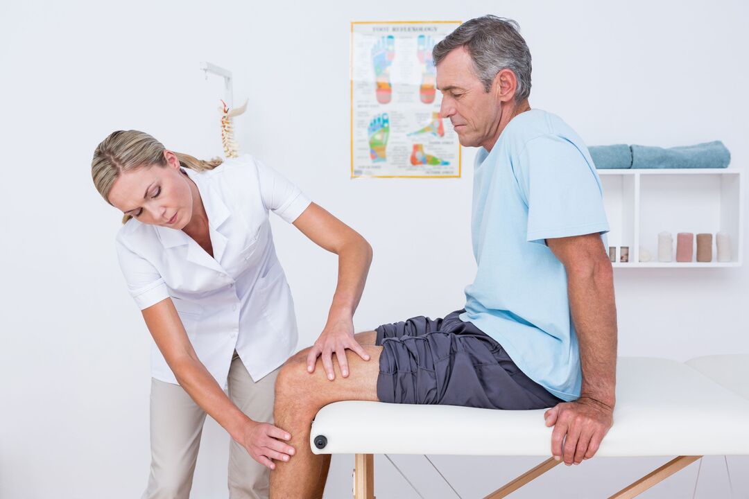
The diagnosis of knee arthropathy is based on the patient's characteristic complaints, changes found during the examination, and the results of other tests.
To confirm osteoarthritis, prescribe:
- X-rays of two projections (anterior and lateral) of the knee joint: the easiest way to confirm the diagnosis in the late stage of pathology;
- Ultrasound: to determine the presence of fluid in the joint, to measure cartilage thickness;
- synovial fluid analysis;
- Diagnostic arthroscopy (visual assessment of cartilage) and biopsy;
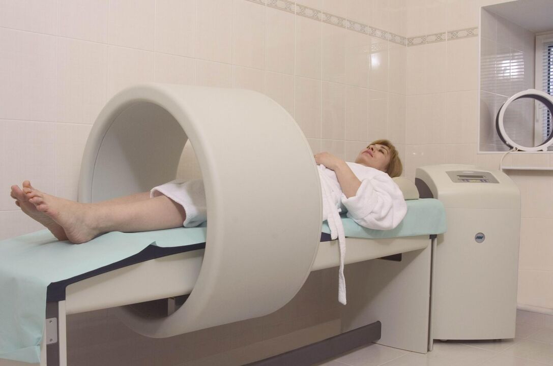
- Computer and Magnetic Resonance Imaging (CT, MRI): Best Methods for Early Diagnosis of DOA.
If a doctor has doubts about the diagnosis, they can prescribe:
- Scintigraphy: scans the joints after the introduction of radioisotopes;
- Thermography: Study of the intensity of infrared radiation (the intensity of which is proportional to the intensity of inflammation).
Treatment of knee joint disease
Treatment options for osteoarthritis combine several approaches: nonpharmacological approaches, pharmacotherapy, and surgical correction. The proportions for each method were determined individually for each patient.
non-drug treatment
In the latest ESCEO (European Society for Clinical Aspects of Osteoporosis and Osteoarthritis) guidelines on how to treat knee osteoarthritis, experts put particular emphasis on patient education and lifestyle changes.
Patients need:
- Explain the nature of the disease and prepare for long-term treatment;
- Teaching how to use assistive devices (canes, orthoses);
- Prescribed diet (for patients with a BMI over 30);
- Perform a set of exercises to strengthen the thigh muscles and reduce the load on the knee joint;
- Explain the importance of increasing physical activity.
In the early stages of knee arthropathy, physical therapy methods have achieved good results:
- massage;
- magnetic therapy;
- Ultra high frequency treatment;
- electrophoresis;
- hydrogen sulfide bath;
- Paraffin application;
- acupuncture.
Medication for Arthritis
Drugs used in DOA are designed to relieve pain, reduce inflammation, and slow the rate of cartilage destruction.
Symptomatic treatment:
- analgesics;
- Nonsteroidal anti-inflammatory substances (NSAIDs) from the COX-2 inhibitor group in tablet or suppository form;
- Non-narcotic analgesics (with resistant pain syndrome).
Structural Modifying Drugs (Chondroprotective Agents):
- chondroitin sulfate;
- Glucosamine sulfate.
These drugs can be taken in capsule form several times a year, injected intramuscularly, or injected directly into the synovial cavity.
Local treatments include proximal and intra-articular injections of glucocorticoids and hyaluronic acid preparations.
An important position in complex treatment in DOA stages I-II is the use of NSAID-based anti-inflammatory ointments, gels, and creams. They help reduce the need for patients to take oral NSAIDs, thereby reducing the risk of damage to the digestive tract.
folk remedies
Topical application of tinctures, decoctions, extracts, medicinal plants should be considered as an adjunct to the treatment of DOA, and folk remedies are not a substitute for those prescribed by doctors.
Plants for osteoarthritis: dandelion, ginger, Jerusalem artichoke, burdock, garlic, sea buckthorn.
Operation
Surgical intervention may be required in all stages of arthropathy in cases where medical measures are insufficient. The most common is endoscopic surgery, which in the most severe cases requires replacement of the endoprosthesis.
Types of Endoscopic Interventions:
- Joint repair and rehabilitation: extraction of inflammatory contents from synovial cavity, cartilage fragments;
- Plasma or laser ablation: removal of mechanical barriers in the synovial cavity;
- Chondrhoplasty.
Corrective periarticular osteotomy is indicated for patients initially presenting with axial limb deformity (no more than 15-20%).
The goal of surgery is to restore the normal shape of the joint, distribute the load evenly over the articular surfaces, and remove the damaged area. This procedure allows you to delay arthroplasty.
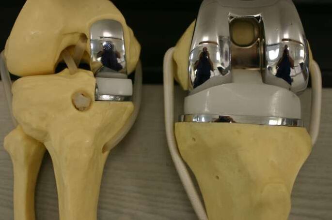
Indications for artificial replacement of the affected area (or the entire joint):
- DOA II-III degrees;
- Severe axial deformity of the limb;
- Aseptic necrosis of subchondral bone layer;
- Persistent pain syndrome.
Contraindications for knee replacement surgery:
- complete damage to the joint;
- unstable ligamentous devices;
- DOA due to inflammatory arthritis;
- Persistent flexion contractures, severe muscle weakness.
In this case, the patient undergoes arthrodesis - a comparison of the knee joint in its physiological position with the articular surface removed. This relieves pain, but shortens the leg, causing secondary damage to the contralateral knee, hip, and spine.
prevention
Prevention of premature cartilage degeneration should begin in childhood.
Precaution:
- Scoliosis prevention;
- Correction of flat feet (shoes with arch support);
- Regular physical education (limiting heavy exercise);
- Exclude fixed postures at work.