


Osteochondrosis is a chronic degenerative dystrophic disease that develops under the influence of many quite different factors. Initially, pathological changes occur in the nucleus pulposus (the inner contents of the intervertebral disc), and subsequently they spread to the annulus fibrosus (the outer shell of the intervertebral disc) and other elements of the spinal motor segment (SDS). This can be the result of the body's natural aging process, or it can occur in the context of injury, increased spinal load, and other causes. In any case, osteochondrosis is only the first stage of disc destruction, and if left untreated, protrusions and hernias develop, which often require surgical removal.
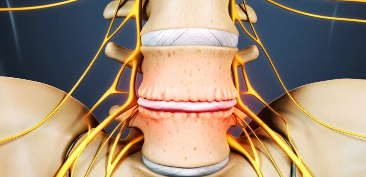
The intervertebral disc is a cartilaginous structure that separates the vertebral bodies and acts as a shock absorber.
Lumbar Osteochondrosis: What It Is
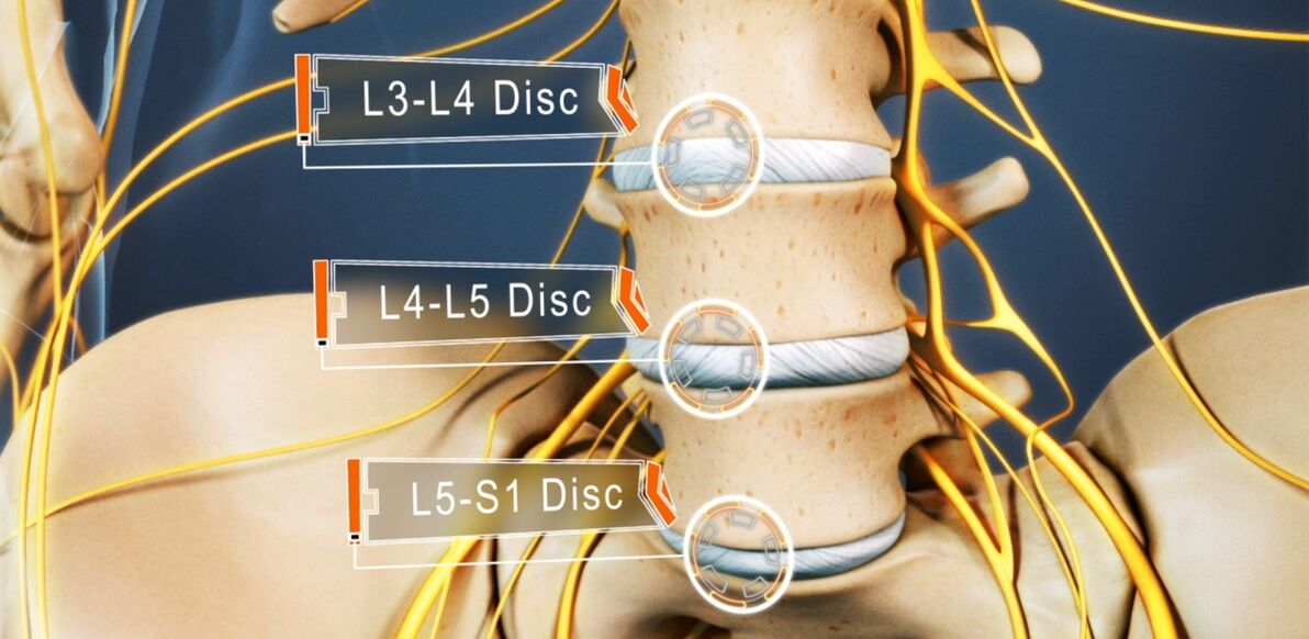
Between 48% and 52% have osteochondrosis. Lumbar osteochondrosis is the most common. The disease can affect any disc in the lumbosacral spine, several, or even all of them. Most of the time, disks L5-S1, L4-L5 are affected, and less so L3-L4. The upper lumbar discs (L3-L2 and L2-L1) are much less affected.
The prevalence of lumbar osteochondrosis is due to the fact that the greatest load of any physical work, especially lifting and carrying weights, walking, running, and sitting all falls on the lower back. The lumbar spine consists of 5 vertebrae and is much larger than the thoracic and cervical vertebrae. Therefore, the discs that separate them are larger in size. Normally, there is a slight lordosis (physiological lordosis) in the lumbar region. It's the last moving part of the spine and it's adjacent to the fixed sacrum, so they're most often talking about lumbosacral osteochondrosis.
If osteochondrosis was previously thought to be an age-related disease, today its first manifestations can already be observed at 15-19 years of age. Already, 1. 1% of 30-year-olds have severe symptoms of degenerative disc dystrophic changes. Among the representatives of the elderly group (over 59 years old), 82. 5% had already developed clinical manifestations of the disease. At the same time, the incidence of pathology continues to increase steadily, largely due not only to the increasing average age of the country's population, but also to changes in lifestyle.
Development reasons
Today, there is still no consensus on the etiology of spinal degenerative diseases. However, the main theory they developed was involution. According to her, osteochondrosis is the result of previous damage to the intervertebral discs and bony structures of the spine, as well as inflammation and other processes that occur. The theory holds that degenerative changes are genetically determined and, in fact, inevitable. Its clinical manifestations, especially in young and middle-aged people, are due to the influence of various endogenous and exogenous factors.
Therefore, the development of lumbar osteochondrosis is facilitated by:
- strenuous physical work, especially in relation to weight lifting;
- a sedentary lifestyle;
- any back injury, including bruises;
- overweight;
- metabolic disorders;
- Violation of posture, deformation of the spine;
- Flat feet and other foot pathologies;
- Pregnancy, especially multiple pregnancies.

onset
Whatever the cause, disc degeneration occurs when the intensity of the catabolic processes (the cleavage and oxidation of molecules) of matrix proteins begins to outpace their rate of formation. One of the key points of this process is the dystrophy of the intervertebral disc.
Since they, like most cartilage in adults, do not have a direct blood supply, as they do not have blood vessels, the supply of nutrients to them and removal of metabolites occurs by diffusion, a process during which the disc moves sequentially with compression and relaxation. The main structure that powers the disk is the end plates on its upper and lower surfaces.
The endplate itself is a bilayer formed by cartilage and bone tissue cells. Thus, the cartilaginous side is adjacent to the disc, while the bone is adjacent to the vertebral body. They are characterized by a sufficiently good permeability, which ensures the exchange of material between cells, the intercellular material of the disc and the blood vessels that pass through the vertebral body. Over the years, especially under the unfavorable influence of internal and external factors, the structure of the endplate changes and the blood supply decreases, resulting in a decrease in the metabolic strength of the intervertebral disc. As a result, its ability to generate new substrates decreases, causing its density to gradually decrease with age.
At the molecular level, this is accompanied by:
- Reduced diffusion rates of nutrients and metabolites;
- decreased cell viability;
- accumulation of cellular decay products and altered matrix molecules;
- Reduce the production of proteoglycans (polymeric compounds responsible for the formation of new chondrocytes, the main source of chondroitin sulfate synthesis);
- Collagen scaffold damage.
possible consequences
Due to the ongoing changes, the disc becomes dehydrated and the nucleus pulposus loses its ability to adequately distribute the load that falls on it. As a result, the pressure within the disc becomes uneven, overloading and compressing multiple annulus fibrosus. Since this happens with every movement a person makes, the annulus is often subjected to mechanical stress. This led to its unfavorable changes.
In addition, reductions in disc height and elasticity often lead to compensatory changes in adjacent vertebral bodies. Bone growths called osteophytes form on its surface. They tend to increase in size over time and even merge with each other, ruling out the possibility of affected PDS movement.
As malnutrition causes damage to the collagen skeleton, the normal structure of the fibers forming the annulus is disrupted under the influence of nucleus pulposus pressure at certain points. Without intervention, this can eventually cause them to crack and break. Gradually, more and more of the annulus is torn where the pressure is applied, causing it to protrude. Increasing the load on the spine especially helps with this. And since the lower back bears the main load during exercise and any physical activity, it is most often affected.
A herniated disc, in which the annulus fibrosus is not eventually ruptured, is larger in size at the base than the herniated part, called a herniation. An intervertebral hernia is diagnosed when it ruptures completely in one place or another.
As part of the fibers in the annulus fibrosus are destroyed, the pressure within the disc gradually decreases, resulting in a decrease in tension and the fibers themselves. This can lead to disruption of its fixation, resulting in pathological mobility of the affected spinal motion segment.
The vertebral body motion segment (SMS) is the structural and functional unit of the spine, consisting of the intervertebral discs, adjacent vertebral bodies, their facet joints, ligaments, and the muscles that attach to these bony structures.
The normal function of the spine can only be achieved if the PDS is properly operated.
Symptoms of lumbar osteochondrosis
The disease can be asymptomatic for a long time, and then begins with mild discomfort in the lower back, which gradually increases. In some cases, however, lumbar osteochondrosis begins acutely, triggering an immediate, intense pain syndrome. In most cases, the first signs of pathology appear after 35 years.
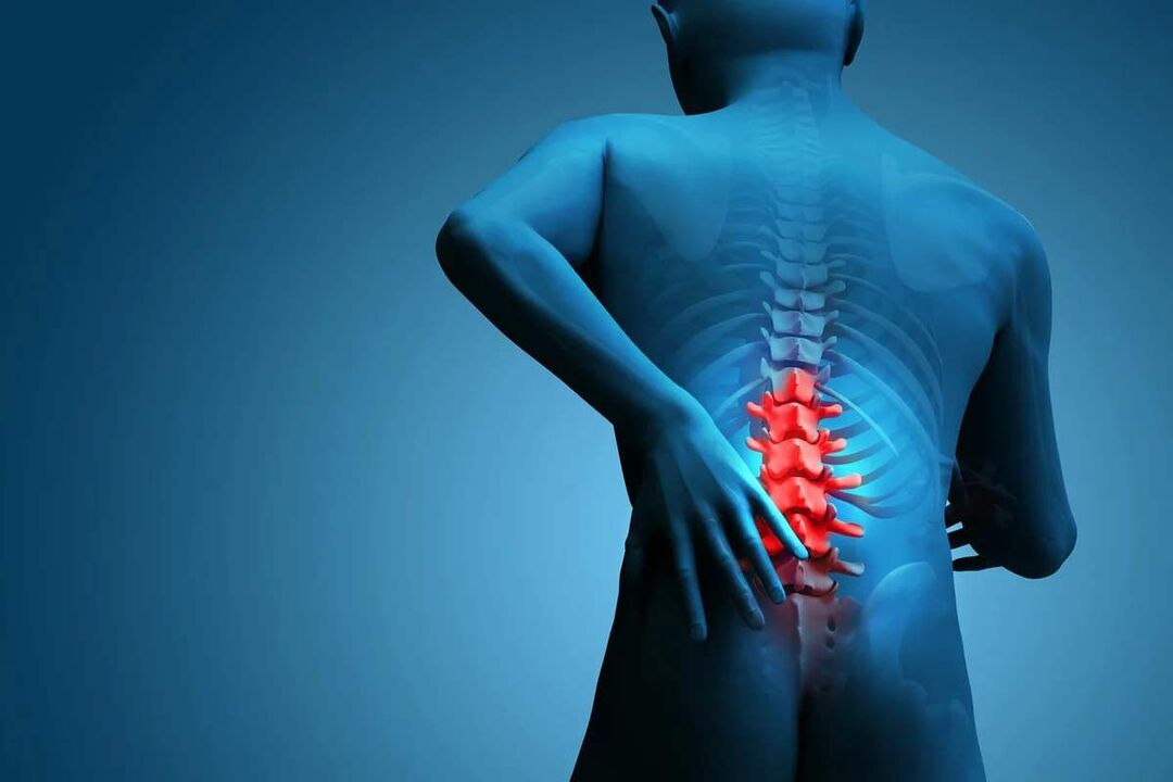
Back pain is the main symptom of the disease. It can vary in character, being painful and tedious, or it can be acute, persistent, or episodic. But basically for pathology, especially in the early stages of development, the alternation of periods of exacerbation and remission is characteristic, and hypothermia or heavy lifting, or unsuccessful sudden movements can all cause another deterioration in well-being.
The pain is usually accompanied by a feeling of numbness and tightness in the back muscles. They are aggravated by physical exertion, sudden movements, heavy lifting, bending over, and even coughing and sneezing.
If, due to the instability of the vertebral body, the nerve roots extending from the spinal cord are pinched by one or the other anatomical structure, this will lead to the development of appropriate neurological disease. Their main manifestations are:
- Shots, severe pain radiating to the sacrum, buttocks, lower extremities, or perineum;
- Sensitivity disorders of varying severity;
- limited mobility, lameness;
- Weakness of muscles innervated by compressed nerves.
In the lumbar spine, the spinal cord ends at the level of 1-2 vertebrae and enters the so-called cauda equina, formed by the accumulation of vertebral roots. Moreover, each of them is not only responsible for the innervation of muscles, but also specific organs of the small pelvis, so prolonged compression can cause the work of the corresponding organs to be disturbed. This can lead to the development of impotence, infertility, gynecological disorders, hemorrhoids and other conditions.
The clinical manifestations of lumbar osteochondrosis, especially in the case of a long disease course and the occurrence of vertebral root compression, are largely dependent on the level of the disease, that is, which particular intervertebral disc has undergone degenerative dystrophic changes.
- L3-L4 Disc Failure - Pain in the front of the inner thigh, calf, and ankle. This is accompanied by decreased sensitivity of the anterior surface of the thigh, decreased severity or loss of knee hops, and decreased quadriceps strength.
- Failure of the intervertebral discs L4-L5 - pain from the upper part of the hip to the outer part of the thigh and calf. Less commonly, this is accompanied by pain spreading to the back of the foot, including 1-3 fingers. In these areas, sensitivity and muscle weakness are reduced. Atrophy and incomplete extension of the big toe sometimes occurs.
- L5-S1 Intervertebral Disc Injury - Pain starts in the mid-buttock area, descends to the heel along the back surface or back surface of the thigh and calf, and can grip the outer edge of the foot, such as 4-5 fingers. In these areas of the lower extremities, sensitivity is reduced, and the gastrocnemius and gluteus maximus are often smaller and accompanied by weakness. If the spinal roots passing at this PDS level are affected, a reduction or loss of Achilles tendon and plantar reflexes may be observed.
The intervertebral discs L1-L2 and L2-L3 are rarely affected.
The pain that accompanies the disease limits a person and significantly reduces his quality of life. Since they persist for a long time and recur periodically, if not always, this cannot but affect the psycho-emotional state. As a result, more than half of the patients showed symptoms such as chronic emotional stress, depression, etc.
diagnosis
If there are signs of lumbar osteochondrosis, you should contact a neurologist or chiropractor. First, doctors collect medical records, which include elucidating the nature of the chief complaint, the characteristics of the pain, the conditions under which it occurs and relieves, the characteristics of a person's work life, etc.
The second stage of diagnosis performed as part of the first consultation with a doctor is a physical examination. During this time, the doctor will assess the condition of the skin, posture, the depth of the physiological curve of the spine, the presence or absence of curvature, etc. The condition of the muscles surrounding the spine (called the paraspinal muscles) must be evaluated because they are often painful and overtense, which is the body's reflex response to inflammation and discogenic pain.
The neurologist may suspect the presence of lumbar osteochondrosis already based on data obtained during examination and questioning of the patient. However, laboratory and instrumental diagnostic methods are required to exclude possible concomitant pathology, as well as to confirm the diagnosis and to accurately determine the extent of injury, the severity of degenerative disc dystrophic changes, and the involvement of skeletal structures.
laboratory diagnosis
Various analyses were not conclusive in the diagnosis of lumbar osteochondrosis. They are more aimed at assessing the extent of inflammatory processes and the detection of concomitant diseases.
Therefore, they can be assigned:
- UAC;
- operator;
- blood tests for blood sugar levels;
- blood chemistry.
Instrument diagnosis
All patients with suspected lumbar osteochondrosis showed:
- Lumbar spine radiographs in two projections - allows you to determine the structure of the bone structure, detect abnormalities, osteophytes formed, changes in facet joints, etc. ;
- CT - allows you to detect changes in bone structure at an earlier developmental stage than X-rays and to identify indirect signs of osteochondrosis;
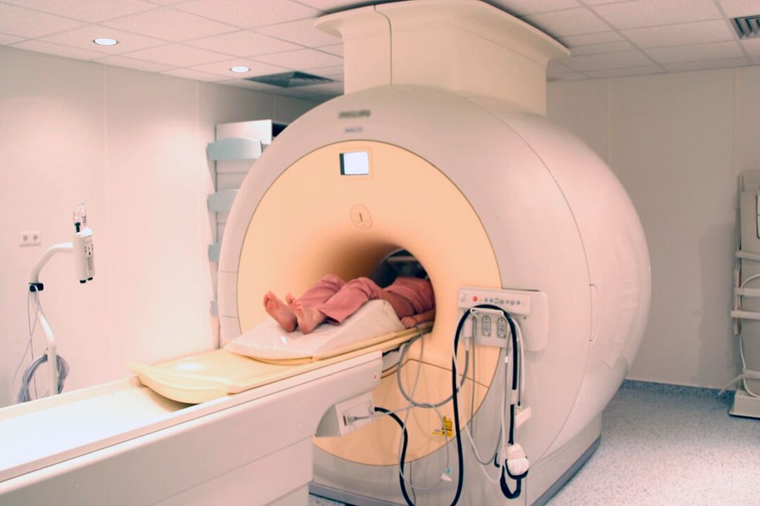
- MRI is the best method for diagnosing pathological changes in chondrogenesis and other soft tissue structures. It can detect the slightest changes in the discs, ligaments, blood vessels and spinal cord and accurately assess their severity and potential risk.
Additionally, it may be suggested that:
- Densitometry - a method of determining bone density, which can diagnose osteoporosis, which is especially common in older adults;
- Myelography - allows you to assess the status of the spinal cord cerebrospinal fluid pathway and the extent of damage to a herniated disc, which is especially important in the presence of a herniated herniation in the lumbar spine.
Treatment of lumbar osteochondrosis
At the time of diagnosis of osteochondrosis, as a rule, all patients are initially treated conservatively, provided there is no apparent and progressive neurological deficit. But her role was chosen strictly individually.
Because the disease is chronic and the disc has an extremely limited ability to regenerate, especially when there are marked degenerative dystrophic changes, the main goal of treatment is to stop its further progression and eliminate the symptoms that plague the patient. Therefore, treatment is always complex and includes:
- medical treatement;
- manual therapy;
- physiotherapy;
- Exercise therapy.
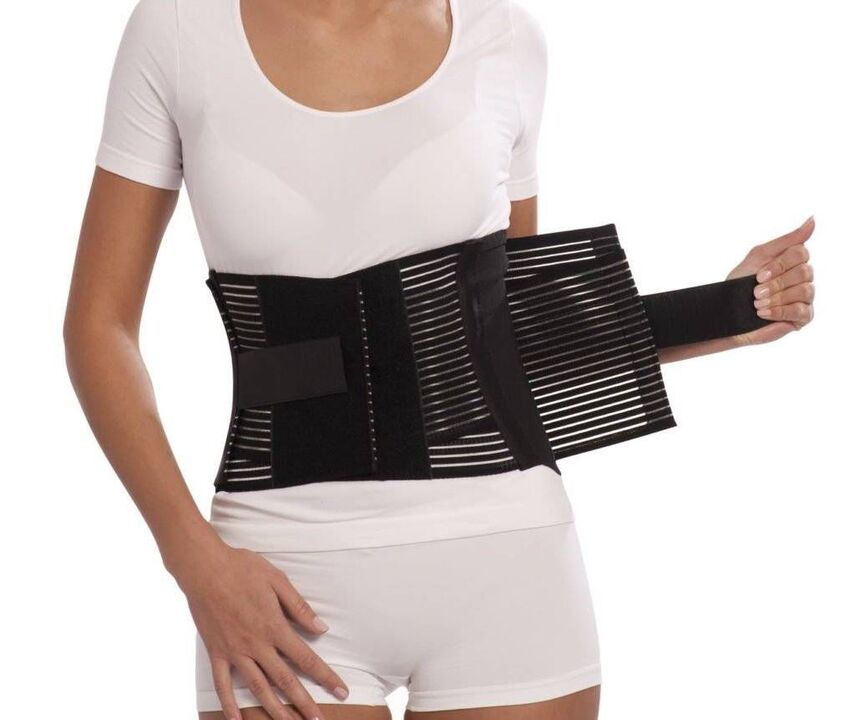
During the acute phase, patients appear to limit physical activity and even insist on bed rest for 1-2 days. This will help relax the muscles and reduce the pressure inside the disc. If you need to sit, walk, or do physical work for long periods of time, you should wear a stabilizing corset.
Instead, after the acute phase and during remission, it is important to move as much as possible, but be careful and rule out increased pressure on the lower back. Patients will need to master the skills of sitting correctly, lifting objects from the floor, and carrying heavy objects, all of which can affect the pathological process. It's important to avoid leaning and sudden movements, and to lift things off the floor or low surface after bending your knees, rather than bending down. You should only sit in a chair that supports your back well, with the back straight. Also, in sedentary jobs, it's important to take regular breaks for short workouts. It is important to avoid falls, jumps, fast running and hypothermia.
For osteochondrosis, it is important to maintain body weight within the optimal range, and for obesity, diet and physical activity appropriate to the patient's condition are required, as excess weight can increase the load on the lower back and lead to a faster progression of pathological changes in the disc.
On average, conservative therapy is usually designed for 1-3 months, although it can last longer. But even after completing the main course prescribed by your doctor, it is necessary to continue taking some medications, exercise therapy and follow lifestyle advice.
medical treatement
The main component of drug therapy is a single drug selected from the group of non-steroidal anti-inflammatory drugs. When choosing them, the doctor must take into account not only the severity of the pain syndrome and the course of the inflammatory process, but also the nature of the concomitant diseases, especially the digestive tract, since long-term use of NSAIDs can adversely affect the state of their mucous membranes and cause digestionExacerbation of various disorders of the system.
It is necessary to treat acute pain in the lower back with NSAIDs and immediately after they occur. Preferably 1-2 days. Depending on the severity of the patient's condition, they can be administered intramuscularly in rectal suppositories, topical and oral forms. Hospital admissions should not exceed 2 weeks. In the future, a separately selected drug will be administered as needed, but frequent use will be avoided.
More recently, drugs as active ingredients have been prioritized more often, among them selective inhibitors of cyclooxygenase-2.
In addition, patients take prescription drugs from the following groups:
- Muscle relaxants - help relax overly tense muscles, thereby reducing back pain;
- chondroprotective agents - the process of improving the metabolic processes of the intervertebral disc (especially effective at the beginning of the earliest stages of the development of lumbar osteochondrosis);
- B vitamins - help improve nerve conduction;
- Antidepressants and anti-anxiety drugs - for long-term osteochondrosis, which causes depression, chronic fatigue, and other psychological disorders.
Therapeutic blockade is used for very severe pain, especially neuropathic pain. They involve the introduction of anesthetics and corticosteroids at the site near the pinched nerve, which quickly relieves the pain. But the procedure should only be performed in healthcare settings by specially trained health workers, as it is associated with the risk of complications.
manual therapy
Manual therapy not only improves the quality of blood circulation in the affected area, but also significantly reduces the severity and duration of osteochondrosis pain. It effectively relieves muscle tension and allows you to eliminate dysfunction, thereby significantly increasing the mobility of affected SMS.
In addition, with good manual therapy, not only can the distance between the vertebrae be increased, returning them to their anatomically correct position, but the compressed nerve roots can also be released. As a result, pain is quickly eliminated and neurological disorders disappear. It also reduces the likelihood of complications and diseases in the work of internal organs.
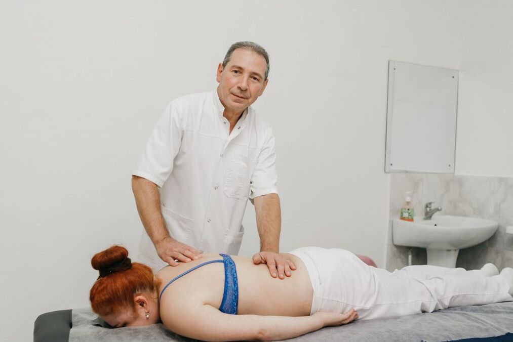
Other positive properties of manual therapy are improved mood, enhanced immunity, activation of the body's natural recovery mechanisms and increased efficiency. There is usually a significant improvement in well-being after the 1st session, and the effect will become more pronounced in the future. Usually, the program consists of 8-15 lessons, and even if the health is fully normalized, it is important to complete it to the end.
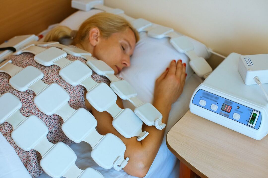
physiotherapy
After acute inflammation subsides, physiotherapy procedures are required, which not only help reduce pain, but also improve repair processes in areas of microcirculation, nutrition, and degenerative dystrophic changes. In most cases, patients are prescribed:
- Electrophoresis of introduced drugs;
- electrical neuromuscular stimulation;
- ultrasound therapy;
- Laser Treatment;
- magnetic therapy;
- UHF.
Which specific physical therapy methods will produce the best results, frequency of delivery, duration of sessions, and likelihood of combination with other types of exposures are determined individually for each patient.
Traction therapy has a good effect on lumbar osteochondrosis. Thanks to it, the distance between the vertebral bodies can be increased, which immediately reduces the load on the affected disc. After the session, to consolidate the results, the patient must wear an orthopedic corset.
exercise therapy
After resolution of acute pain, the treatment regimen must be supplemented with exercise therapy. Its main task is to stretch the spine and relax spastic muscles in the lower back. Additionally, therapeutic exercises help strengthen the muscular corset, provide reliable support for the spine and improve posture. During this process, blood circulation is inevitably activated and metabolic processes are improved, which has a beneficial effect on the nutrition of the intervertebral disc.
For each patient, a separate set of exercises was selected based on the degree of degenerative malnutrition changes, the patient's level of physical development, the nature of concomitant diseases, age, and other factors. Initially, it is recommended to study under the guidance of an experienced sports therapy coach.
It is recommended that all patients with spinal degenerative changes go to the swimming pool 2-3 times a week as swimming lessons minimize the load on the spine but are effective in strengthening the back muscles.
Therefore, lumbar osteochondrosis is one of the most common diseases. At the same time, it can deprive a person of the ability to work for a long time, and even lead to disability due to the development of complications. Therefore, it is important not to ignore the first symptoms of the disease, as it is the easiest to deal with. With the onset of pain, especially numbness, reduced mobility, and back pain, you need to contact a neurologist as soon as possible to get the necessary tests and start treatment. In this case, it is possible to stop the pathological process and return to a normal, fulfilling life without suffering and major limitations.